Japanese scientists have successfully conducted a kidney tissue transplant between rat fetuses while still in their mother's womb. Their study focuses on a rare neonatal condition called Potter Syndrome.
What is Potter Syndrome?
Potter Syndrome is a medical condition that influences how a fetus develops in the mother's womb, affecting the growth and function of a baby's internal organs like kidneys. Also known as Potter Sequence, it affects about 1 in 4,000 to 10,000 births.
This neonatal disorder occurs when there is not enough amniotic fluid that surrounds a fetus as it grows in the uterus. Babies born with this condition usually do not survive long enough to receive dialysis or other forms of treatment.
Because of this challenge, a way must be found to bridge the gap until the babies are old enough to undergo invasive treatments. Scientists believe transplanting fetal pig kidneys into human fetuses can provide the solution, but this is still a highly experimental approach.
Transplanting an organ before a baby is born could allow the tissues to grow and develop with the fetus. This would allow the organ to function at birth with less risk of rejection.
World's First Fetus-to-Fetus Transplant
At Jikei University School of Medicine in Tokyo, Takashi Yokoo led a team of researchers in conducting an experiment that could unlock the future of in-utero xenotransplantation in humans. This could be the first step to transplanting fetal pig kidneys into human fetuses. The details of their study are discussed in the paper "Fetal Kidney Transplantation for In Utero Fetuses."
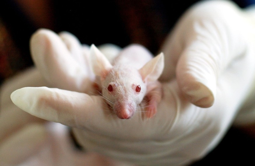
The surgery involved anesthetizing pregnant rats and exposing their uterus carefully. The tiny kidneys which have been removed from the donor fetuses were injected through the uterine wall of the recipient fetus. The transplanted kidneys were labeled with a green fluorescent protein to enable the surgeons to check if the tissue had been successfully transplanted.
The procedure's success rate was 88%. Of the nine fetuses, only one did not show evidence of the green fluorescent protein when they were born a few days after the surgery. According to Yokoo, the fetus that did not develop fluorescent-green kidneys probably did not transplant the tissue successfully.
The kidneys also seemed to develop normally, although they had to be drained manually since they separated from the animals' urinary tracts. Yokoo and colleagues noted that the blood vessels of the host rat started to grow inside the donor tissue. This is a good thing regarding the likelihood of transplant rejection.
The team also experimented with fetal mouse kidneys and confirmed the maturation of the transplanted kidney tissues. They demonstrated less tissue damage from rejection compared to the transplantation of mouse fetal kidneys into adult rats.
The research team plans to apply for ethical approval to perform their experiments in humans. They also start engaging with the public to inform them of the benefits of human fetal xenotransplantation. They are hopeful that in-utero transplants could be a game-changer for babies born with various kinds of organ defects.
RELATED ARTICLE : Pig Heart Transplant Performed For the 2nd Time in the World With Hopes of Prolonging Dying Man's Life
Check out more news and information on Xenotransplants in Science Times.
© 2026 ScienceTimes.com All rights reserved. Do not reproduce without permission. The window to the world of Science Times.


