Every year since 1975, camera manufacturing giant Nikon has held the Nikon Small World Competition to recognize the art in science through photography using a light microscope. This year's event has been recently concluded, and the winners have been announced.
The Top Placers
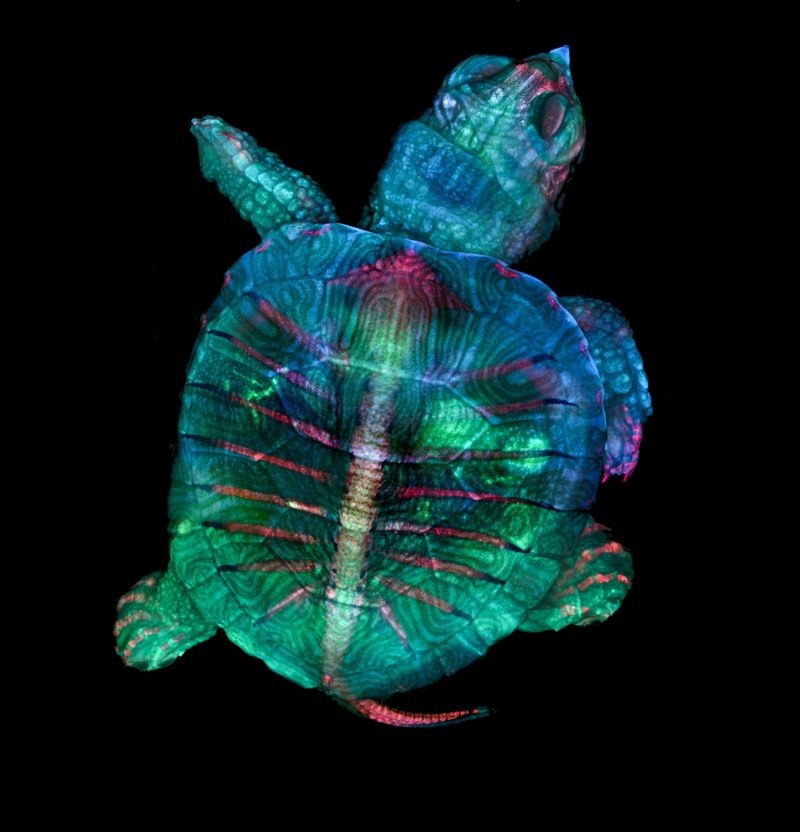
Taking first place is a fluorescent turtle embryo taken by microscopy technician Teresa Zgoda and micro photographer Teresa Kugler. The turtle embryo, being only less than an inch long, required a more intricate photography method as its details could not be captured in a single image. The two then decided to combine hundreds of images, which individually gave focus on different points of the image.
In second place is an image of three stentors. These are single-celled microbes that are usually found in fresh water and are also sometimes called "trumpet animalcules" to give credit to their shape. The artist, Dr. Igor Siwanowicz, is a research scientist at the Janelia Research Campus at the Howard Hughes Medical Institute. His image of the trumpet-shaped creatures presented the cilia—the hair-like part of the microbe's body used for its movement and feeding—in vibrant colors against a black background.
But the real challenge in capturing the image was thinking of how to keep the microorganisms still. Being the tiny microbes that they are, stentors are constantly moving. Siwanowicz then thought to expose them to magnesium ions, which did the trick. "Think a relaxing Epsom salt bath," Siwanowicz simply explained.

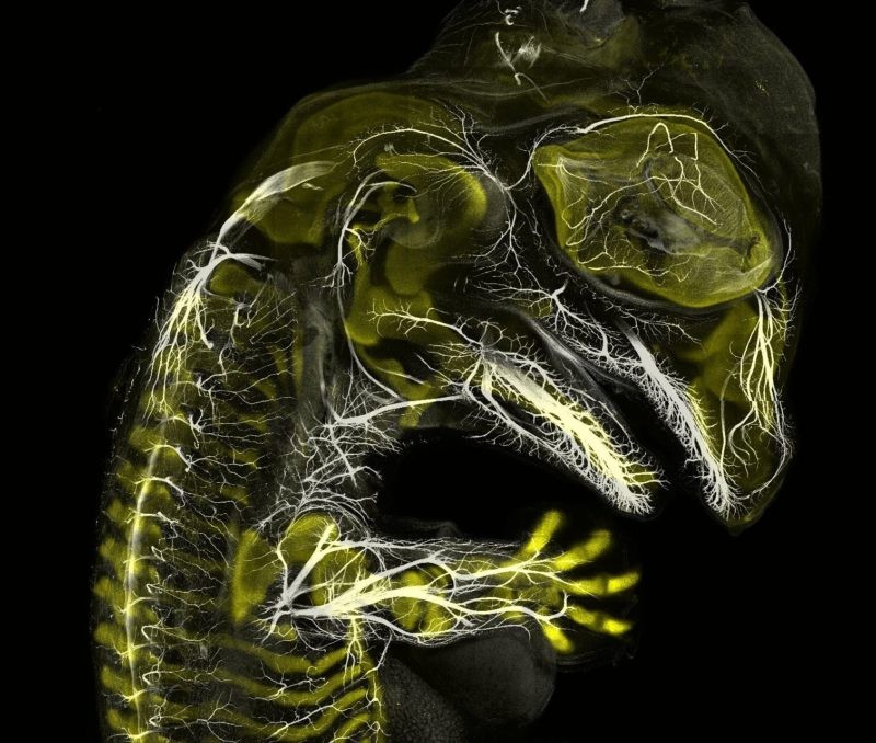
Hitching third place is a 20-day-old American alligator embryo. Yale University paleontology and development lab graduate student Daniel Smith Paredes used various immunological stains to be able to show the nerves of the reptile in white hue against the developing bones in yellow. Like the turtle embryo image, this entry is a combination of a number of pictures. Paredes now continues to study the different stages in the development of a variety of vertebrates.
Aside from these three stunning photos, 17 others were recognized in Nikon's website to form the top 20.
Honorable Mentions and Images of Distinction
Aside from the 20, Nikon also gave Honorable Mentions to 15 entries, among which are Nathan Burns' take on the endothelial cells of the intestine of a mouse embryo, Caleb Dawson's image of the myoepithelial cells of a lactating mouse, and Dr. Tagide deCarvalho's image of penicillium mold spores.
The manufacturing giant also gave recognition to 62 Images of Distinction. Some of these are Christopher Algar's Phantom midge larva, Langley Anderson's crepe myrtle, and Dr. Haris Antonopoulos's image of a bearing from a mechanical watch.
Nikon Continues to Inspire
In parallel with the photography competition is Small World In Motion, the company's video competition where the movie or time-lapse entries involving the use of a microscope are deliberated. And although the 2019 competitions have already been concluded, the company will be collecting entries for next year's competition soon.
© 2026 ScienceTimes.com All rights reserved. Do not reproduce without permission. The window to the world of Science Times.


