A conventional light-based lets us observe specimens for up to 200 nanometers. Everything that is closer or lesser to the figure will be impossible to distinguish. But recently, University of California San Diego's engineers have identified a smart way to take the conventional microscope's resolution to the next level. Amazingly, the upgrade requires no additional equipment or lenses that should be attached to the microscope.
Rayleigh Criterion and the Limits of Resolution in Traditional Microscopes
Rayleigh Criterion is an 1896 theory created by John William Strutt, 3rd Baron Rayleigh that tackles the limits of resolution. Based on the theory, the resolution of a typical light-based microscope is affected by the optical function of the glass lenses that will be attached to it, as well as the characteristics of the light source itself.
Light rays play a great part in increasing resolution. It produces diffraction that allows us to perceive samples on a microscope, making them appear closer.
Electron microscopes, on the other hand, fires off a beam of electrons instead of visible light. This kind of microscope is so powerful that it can be used to observe a specimen lesser than a single nanometer. Yet, there is a catch in using electron microscopes. Living subjects that are place in the vacuum chamber of the microscope are being killed by the electron blast. This limits the research for cells and other living organisms. With that said, there are no alternatives to a high-resolution microscope until now.
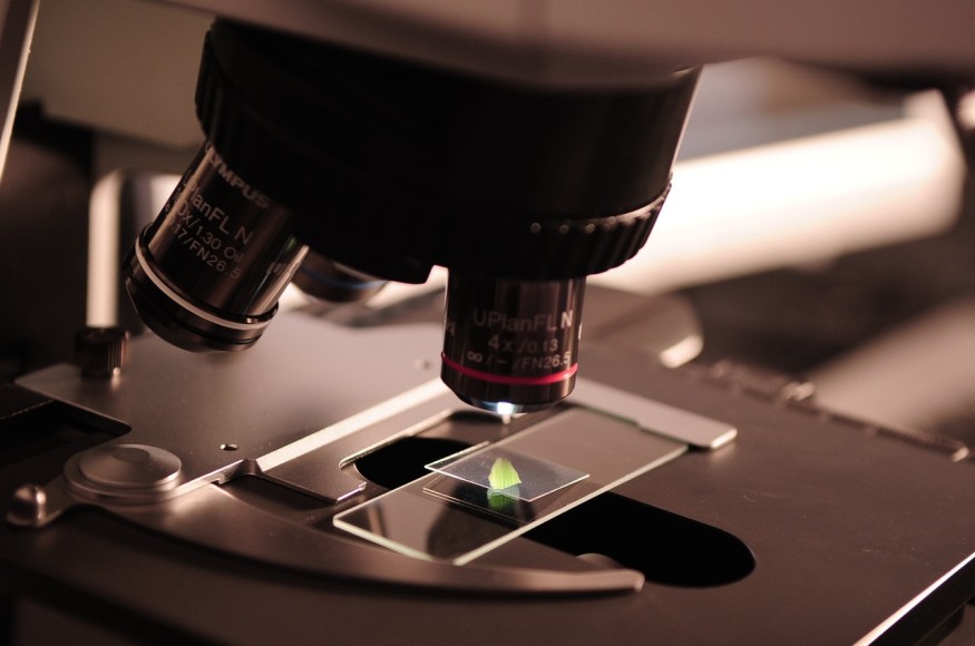
Observation of High-Resolution Subjects Possible with Light Shrinking Slide Boost
UCSD engineers recently created a high-resolution microscope that did not necessarily demand any fancy upgrades and additional hardware. They developed a hyperbolic metamaterial that has unique structures which are used to manipulate light. Hyperbolic materials are initially applied for improving optical imaging. The engineers unutilized the material to the microscopic slide where the specimen is placed reports Gizmodo.
Hyperbolic metamaterials used for producing high-quality images are made from alternating silver and silica glass that measures nano-thin. The wavelengths of the visible light will shorten as it passes through the super-thin sheets of the metamaterial, resulting in speckled patterns.
The speckled light patterns will help the sample to be more visible as it illuminated the microscope slide from all directions. It will highlight each angle of the low-resolution images captured, that once assembled, will be optimized through a reconstruction program. The program will then put together the gathered images to form a high-resolution image.
The 'light shrinking slide boost' technology's process and information are published in Nature Communications entitled "Metamaterial assisted illumination nanoscopy via random super-resolution speckles." Overall, the possibility of observing the known tiniest specimens is at hand through the use of conventional microscopes. Resolution-wise, the electron microscope is still faster at producing high-resolution images of the subjects.
But compared to the electron-blasting device, the light-based microscopes are still better, as it allows the normal 40 nanometers of resolution to be used without pulverizing the living specimen. With the recent discovery, the affordability and safety of the conventional microscope will undoubtedly improve laboratory activities. The traditional device has opened a new route to bio-imaging and other future studies of smaller scale organisms.
RELATED ARTICLE : "Game-Changing" Nanomaterials Are Both Generators and Sensors; Here's How They Work
Check out more news and information on Technology & Innovation on Science Times.
© 2026 ScienceTimes.com All rights reserved. Do not reproduce without permission. The window to the world of Science Times.


