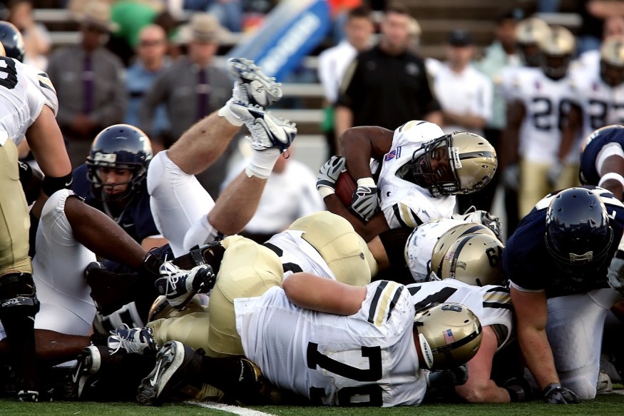
The magnetic resonance imaging (MRI) scans on athletes who played more contact sports, such as American football, showed lesions to the brain's white matter, called white matter hyperintensities.
The recent study titled, "Association Between Antemortem FLAIR White Matter Hyperintensities and Neuropathology in Brain Donors Exposed to Repetitive Head Impacts" published in the medical journal of the American Academy of Neurology, revealed that the more years they play, the more white matter brain lesions they have that are associated with neuropathological changes.
Routine MRI Spots Signs of Sports-Inflicted Neurological Damage
Repetitive head hits are common in contact sports, such as American football. However, the extent of their brain damage is not usually studied not until their autopsy, which is done when they are already dead.
US News reported that the new study suggests that spotting any signs of sport-inflicted neurological damage can be done even without waiting for an athlete to die. The research shows that routine brain scans can reliably spot these signs and that the more brain lesions there are, the longer a football player has been playing the sport.
Study author Michael Alosco, the co-director of the Alzheimer's Disease Research Center Clinical Core under Boston University's School of Medicine, said that MRI scans might be able to capture long-term harm to the brain in people exposed to repetitive hits to the head, especially those who play contact sports.
Together with his team, they explained that MRI looks for the white matter hyperintensities that appear as bright white spots that anyone can see them. These lesions to the brain's white matter signal injury associated with contact sports.
Alosco noted that outside sports, these lesions can be observed in people older than 65 with heart disease as the root cause. When the heart fails to pump oxygenated blood to the brain, it results in an oxygen deficit that injures the person's small blood vessels and white matter.
Effects of Repetitive Head Impacts On An Athlete's Brain
The team set out to see the effects of repetitive head impacts on an athlete's brain. According to the press release, the study involved 75 people who experienced repetitive head impacts and reported head injury symptoms.
About 67 of these participants were football players and eight of them were athletes of other contact sports, such as boxing and soccer. Each football player played an average of 12 years of which 16 have played professionally and 11 were semi-professional football players.
They donated their brains after their death to advance research into the long-term effects of repetitive head impacts. Aside from that, researchers also reviewed brain scans of the participants when they were alive during which when they were 62 years old, on average.
Researchers found that 64% of them had dementia before their death and autopsies showed that 71% had chronic encephalopathy (CTE), a neurodegenerative disease due to repetitive head impacts.
The team noted that brain scans revealed twice the odds of having more severe small vessel disease and lesions on the white matter, and three times more severe tau accumulation in the frontal lobe. Tau accumulation is a biomarker for dementia, Alzheimer's disease, and CTE.
RELATED ARTICLE: Long-Term Consequences of Traumatic Brain Injury
Check out more news and information on Brain in Science Times.
© 2026 ScienceTimes.com All rights reserved. Do not reproduce without permission. The window to the world of Science Times.


