New sensors may provide doctors and cancer researchers with new capabilities by examining enzyme activity at the organism, tissue, and cellular sizes.
The "hallmarks of cancer" are essential molecular changes in cancer cells and their surrounding tissue to enable tumor formation, growth, and survival.
While certain genes and proteins cooperate with other cellular functions cause these alterations, the details differ depending on the kind of cancer and even from patient to patient.
Phys.org said researchers have gained an understanding of the many cell types in a tumor's microenvironment and how its composition varies in response to therapies thanks to sensitive methods for analyzing protein or gene expression, even at the single-cell level.
However, these tests don't always reveal which proteins are active or crucial to tumor progression, nor do they necessarily enable doctors to noninvasively track the development of a patient's condition or how well they respond to therapy.
For instance, a protein could be a passive observer rather than an active participant in a cancer cell's biological alterations. Enzymes may clarify which genes or proteins to target at a specific moment since they catalyze biochemical events inside cells.
Nanosensors Will Monitor, Examine Cancer Cells
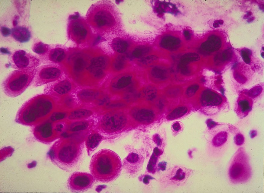
Researchers from the MIT Koch Institute for Integrative Cancer Research have created a set of enzyme-targeting nanoscale tools to monitor cancer progression and treatment response in real time, map enzyme activity to specific locations within a tumor, and isolate pertinent cell populations for analysis. Their findings were recently published in Nature Communications.
Senior study author Sangeeta Bhatia, the John J. and Dorothy Wilson Professor of Health Sciences and Technology, professor of electrical engineering the computer science, said they anticipate that this new set of instruments will benefit the lab and the clinic.
"With further development, the nanosensors could be used by clinicians to tailor treatments to a patient's specific cancer, and to monitor cancer progression and treatment response, while researchers could use them to better understand the molecular biology of cancer and develop new tools to diagnose, track, and treat the disease," said in a MIT News report.
The Bhatia laboratory has been creating noninvasive urine tests for cancer diagnosis for several years, including lung, colon, and ovarian cancer. Nanoparticles interacting with tumor proteins, known as proteases, are the basis for the testing. Proteases are a class of enzymes that cut proteins into smaller pieces by acting as molecular scissors. Proteases break apart the extracellular protein web that binds cells in place, allowing cancer cells to escape from tumors.
Peptides are condensed protein fragments that target cancer-related proteases, are coated on the nanoparticles. The peptides are cleaved when the nanoparticles reach the tumor location, releasing biomarkers that may be seen in the urine.
The goal of the present study was to see if this technology might be used to correctly and sensitively follow the progression of cancer over time and how it responds to various therapies. To target the overexpressed proteases in a mouse model of non-small cell lung cancer, the scientists developed a panel of 14 nanoparticles. When these nanoparticles come into contact with improperly functioning enzymes in the tumor microenvironment, they are modified to release barcoded peptides.
Each nanosensor was able to monitor various protease activity patterns, which sharply varied as the tumor grew. Within three days of delivering a lung cancer-targeting medication, the researchers identified early evidence of tumor regression.
Although experts could use the already available nanosensor method to monitor tumor development and treatment response generally, it was unable to provide any insight into the precise biological mechanism at play.
The researchers aim to pinpoint the precise protease active in pericytes and analyze its function in angiogenesis. They intend to use this information to create therapeutic formulations that may be administered to patients to prevent the recruitment and creation of blood vessels linked to tumor growth.
RELATED ARTICLE : New AI Technology Detects Invisible Patterns of MRI to Accurately Predict If and When a Person Dies From Cardiac Arrest, Study Claims
Check out more news and information on Artificial Intelligence in Science Times.
© 2026 ScienceTimes.com All rights reserved. Do not reproduce without permission. The window to the world of Science Times.


