A new study establishes virtual imaging trials as a guide for effectively assessing and optimizing computerized tomography (CT) and radiography analysis tools to be used against the global coronavirus disease (COVID-19) pandemic.
Through an open-access article in the American Journal of Roentgenology (AJR), the monthly peer-reviewed journal published by the American Roentgen Ray Society (ARRS), the authors of the study details the first computational models created from confirmed COVID-19 cases. Using the developed models as their proof of principles, the researchers suggest that these models can be used to guide imaging simulators in future coronavirus imaging studies.
RELATED: Hong Kong Scientists Report Their First COVID-19 Reinfection
Virtual Imaging Trials for COVID-19 Studies
The open-access report notes that virtual imaging trials have two main components. One is the representative models of targeted subjects while the other refers to realistic models of imaging scanners. Additionally, in developing their COVID-19 computational models, researchers identified three requirements: modeling the body habitus, or the build, modeling the specific morphologic features related to the abnormality, and modeling both the material and texture of the required abnormality.
For the body habitus, researchers used existing computation models - specifically the 4D extended cardiac-torso (XCAT) model developed by radiologists at Duke University. The choice was made because of the XCAT phantom's representation of both sexes at a combination of varying ages, heights, and weights - derived from actual patient data.
In terms of the abnormality's morphologic features, researchers focused on the most common CT manifestations for the coronavirus disease - ground-glass opacities (GGO). This refers to the patterns that make COVID-19 infected lungs appear lighter or grayish, as opposed to the black CT scan results for normal lungs. After getting the necessary approvals, researchers collected and analyzed 20 confirmed positive cases of COVID-19.
Lastly, for the texture and material requirement, authors of the study modified existing models of the underlying lung parenchyma - the functional lung tissues, opposite of connective and supporting tissues - to match observed properties of those with the coronavirus disease.
RELATED: AeroNabs: A Novel Protection Against COVID
For CT and Radiography Simulation
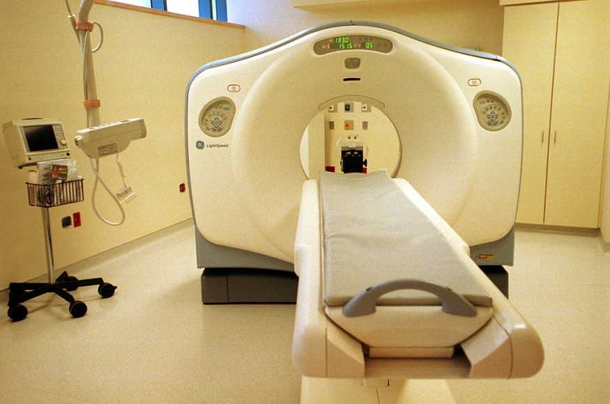
The simulation models used in the virtual imaging trials are those previously reviewed by the researchers in an earlier report. They used DukeSim, a hybrid simulator that uses the ray-tracing and Monte Carlo simulation techniques in computer graphic simulations, to generate its radiographic images. They then used the Definition Flash from Siemens Healthineers as their CT scanner, validated through DukeSim, to test out their developed models. With the results, they were able to show that chest CT scans and radiography are promising modalities for detecting COVID-19. Based on the comparative scans, researchers concluded that the simulated abnormalities were realistic - shape and texture-wise.
With this, the authors of the study suggest that the toolsets they developed will be able to lead the use of virtual imaging trials to assess and optimize these existing tools in the coronavirus pandemic efforts. Through the models they developed, virtual imaging trials could support a comparison of chest radiography and CT for patient screening; another application is in determining the effects of radiation dosage of detection accuracy, better using this specific parameter to better image COVID-19 patients.
Check out more news and information on Xray on Science Times.
© 2026 ScienceTimes.com All rights reserved. Do not reproduce without permission. The window to the world of Science Times.


