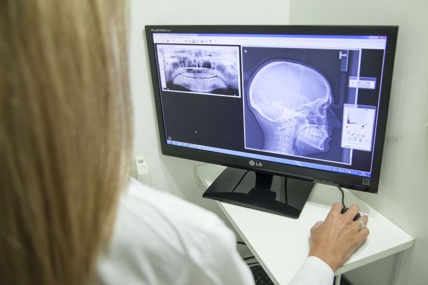
Scientists from the ARC Center of Excellence in Exciton Science, based at Monash University and RMIT University, have used tin mono-sulfide (SnS) nanosheets to develop the world's thinnest X-ray detector that has the potential to enable real-time imaging of cells in the body someday.
SnS nanosheets are used in photovoltaics, field-effect transistors, and catalysis, showing great promise. This time, the team has shown that they are also excellent candidates for soft X-ray detectors.

World's Thinnest X-Ray Detector
In the study, titled "Soft X-ray Detectors Based on SnS Nanosheets for the Water Window Region" published in the journal Advanced Functional Materials, researchers indicated that SnS nanosheets possess high photon absorption coefficients that enabled them to create ultrathin soft X-ray detectors that are highly sensitive and has a rapid response time.
Phys.org reported that the materials the team used were found to be even more sensitive than the metal halide perovskite, another emerging candidate, and boasts a faster response time than known detectors. More so, the sensitivity of these materials can be adjusted across the soft X-ray region.
The world's thinnest X-ray detector is less than 10 nanometers thick or ten times thinner than a sheet of paper about 100,000 nanometers thick. The last thinnest X-ray detectors were between 20 and 50 nanometers, which means this new one has broken the record.
However, researchers said that considerable work is still needed to explore its full potential. They noted that SnS nanosheets respond within milliseconds, which allows them to scan something and get an image almost instantaneously. In theory, it could allow the doctor to see results in real-time due to its high sensitivity and high time resolution.
It can also be used to see cells interact with each other, such as seeing proteins and cells evolve and move using X-rays.
ALSO READ: Researchers Fabricate Novel X-Ray Photodetectors by Embedding Perovskites on Graphene
Why is a Highly Sensitive and Highly Responsive X-ray Detector Important?
According to Science Daily, there are two types of X-rays: hard X-rays for scanning broken bones and other illnesses; and soft X-rays used to study wet proteins and living cells.
Some x-ray measurements take place in a water window or the region of the electromagnetic spectrum wherein water is transparent to soft X-rays. Typically, soft X-ray detections are done using the hugely expensive Synchrotron. However, recent advances in on-synchrotron soft X-ray laser sources have lower cost and portable designs that provide an accessible alternative for researchers.
But researchers still need soft X-ray detector materials that are highly sensitive to low-energy X-rays because direct detection is better, easier to prepare, and thinner than indirect approaches.
Dr. Nasir Mahmood of RMIT University, one of the study's lead authors, said that the sensitivity and efficiency of SnS nanosheets depend on how thick they are and their lateral dimensions, which are impossible to control.
So, scientists thought of using a liquid metal-based exfoliation method to produce high-quality large-area sheets with a controlled thickness that efficiently detect soft X-ray photons in the water window. They stacked ultrathin layers to further improve their sensitivity, which is now better than existing direct soft X-ray detectors.
RELATED ARTICLE : New X-Ray Scanner Instantly Detects an Object's Molecular Composition
Check out more news and information on X-Rays in Science Times.
© 2026 ScienceTimes.com All rights reserved. Do not reproduce without permission. The window to the world of Science Times.


