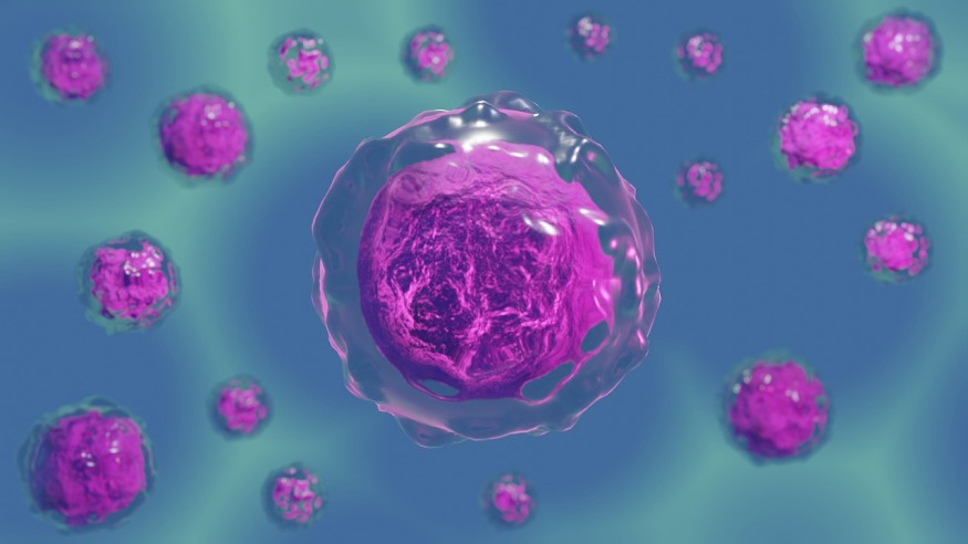
Cranial neural crest cells (CNCCs) form many body parts of vertebrates from fish to humans and generate tissues. A press release from the University of Southern California (USC) reported that scientists from the lab of Gage Crump have sought to understand the versatility of these stem cells, so they created a series of atlases over time to comprehend better how CNCCs work at the molecular level.
Stem cell biology and regenerative professor Gage Crump from the Keck School of Medicine of the USC said that CNCCs have long fascinated biologists, and through studying its process, they could finally identify the switches that allow CNCCs to form into different cell types and gain insights on head development and craniofacial birth defects.

What Cranial Neural Crest Cells (CNCCs)?
Cranial neural crest cells (CNCCs) are derived from the ectoderm and sometimes referred to as the fourth germ layer because of their major importance, according to the 6th edition of the book Developmental Biology.
These stem cells migrate extensively to generate many differentiated cell types, such as neurons and glial cells of the nervous system, the epinephrine-producing cells of the adrenal gland, the pigment-containing cells in the epidermis, and the skeletal connective tissues in the head.
An article in Science Direct reported that CNCCs contribute much to the formation of the bones, cartilage, and many parts of the head and teeth. Any defects to the development of these cells could cause craniofacial deformations.
The interactions between CNCCs, facial epithelium, and neuroectoderm have contributed to the development of ost craniofacial structures in vertebrates. However, different species have different facial morphologies.
Tracking CNCCs to Generate Sufficient Data for the New Computational Tool
Researchers of the new study, titled "Lifelong Single-Cell Profiling of Cranial Neural Crest Diversification in Zebrafish," published in the journal Nature Communications, labeled CNCCs in the zebrafish with a red fluorescent protein to keep track of which cell types came from them throughout their lifetime.
According to Phys.org, the team employed a single-cell genomics approach to determine the complete set of active genes and the organization of the DNA across hundreds of thousands of individual CNCCs. Then they developed a new computational tool to make sense of their data.
They called their new computational analysis "Constellations" because the final visual output of their approach yields a reminiscent of constellations of stars in the sky. Although the Constellations algorithm can predict the future of the cells and which genes control their development, unlike the constellations in the sky.
Researchers discovered that these stem cells do not initially have all the necessary information to make many cell types. They only gain it upon dispersal throughout the embryo of the fish when they reorganize themselves as they prepare to become specialized tissues.
The real-life application of the Constellations algorithm reveals that it can accurately pinpoint the role of a family of "FOX" genes in facial cartilage formation and the GATA genes as a powerful tool in studying organ system development and regeneration.
RELATED ARTICLE : Origin of Life Allegedly Driven By Organisms From Outer Space
Check out more news and information on Biology in Science Times.
© 2026 ScienceTimes.com All rights reserved. Do not reproduce without permission. The window to the world of Science Times.


