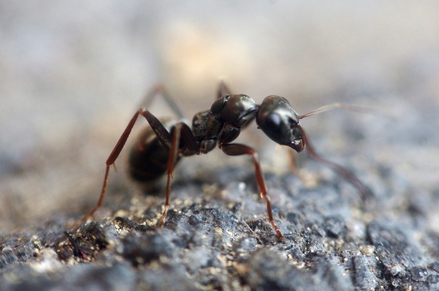
Lithuanian photographer Dr. Eugenijus Kavaliauskas snapped the ant magnified five times under a microscope, revealing its red eyes and shadowy face in exceptional detail.
Nikon's Small World Photomicrography Competition
The photo, according to Daily Mail, was submitted in the Nikon Small World Photomicrography Competition by Kavaliauskas. It is one of the 57 Images of Distinction.
According to Eric Flem, Communications and CRM Manager at Nikon Instruments, Nikon Small World receives a variety of microscopic photos each year that showcase exceptional scientific skill and aesthetics. This year, he claimed, was no different.
The competition this year features breathtaking images created by scientists, artists, and photomicrographers from all over the world, with various degrees of experience and educational backgrounds.
Captured Ant Image
The photographer described to Insider how he managed to capture the ant in a woodland close to his Taurag, Lithuania, house. He said that the basic objective of photography is to be a discoverer. He continued that he is constantly searching for details, shadows, and unknown corners. He expressed his fascination with God's creations and the chance to witness them.
Terrifying Photo
Despite the fact that the ant could seem somewhat frightening, Kavaliauskas is confident that nature is free of dangers.He admitted that when he first began using microphotography, he, too, believed that all beetles somewhat resembled monsters. But he's gotten used to it now. He was taken aback by the sheer number of intriguing, gorgeous, and little-known miracles that exist beneath our feet.
According to his portfolio, Kavaliauskas has received several photography awards for his birds of prey photos.
Images of Distinction
The photograph by Kavaliauskas wasn't the only close-up of an insect that Nikon showcased.
The Images of Distinction category also included a stunning image of a red speckled gem beetle and a vibrant image of a fearless jumping spider.
However, Grigorii Timin took first place this year for his incredible photograph of a Madagascar red-spotted day gecko's developing hand.
The embryonic hand, according to Timin, is roughly 3 mm (0.12 in) long, making it a sizeable sample for high-resolution microscopy. The scan takes more than two days to acquire and generates roughly 200 GB of data because it comprises 300 tiles, each of which has about 250 optical sections.
Caleb Dawson received the second prize for his breast tissue photograph that showed contractile myoepithelial cells encircling milk-producing alveoli.
The myoepithelial cells were imaged using a confocal microscope after being dyed with several fluorescent dyes over the course of a week.
Satu Paavonsalo and Dr. Sinem Karaman won third place for their photograph showing the blood vessel networks in an adult mouse's intestine.
ALSO READ: Ant Teeth: Scientists Reveal How They Can Help in the Development of Gadgets
Photomicrography
Traditional microscopy methods, such as bright field and cross-polarized illumination, are used as the foundation of photomicrography. The majority of microscopes that are used in biological laboratories use transmitted light and function in bright field mode. The amount of transmitted light is reduced to around 30% of the emitted value when a polarizing device is introduced into the light path.
Two separate polarizing elements-one called a polarizer and the other an analyzer-are put into the light path with their vector proportion planes crossed at a 90° angle with respect to one another in order to produce cross-polarized illumination from a bright field microscope.
Check out more news and information on Technology in Science Times.
© 2026 ScienceTimes.com All rights reserved. Do not reproduce without permission. The window to the world of Science Times.


