Scientists were able to grow full human embryo models in the lab. These models are observed to depict features that typical two-week-old embryos possess.
Studying Human Embryonic Development in an Ethical and Accessible Way
The embryos were grown out of lab-cultured stem cells. These models are known as stem cell-based embryo-like structures (SEMs). They were nurtured without eggs, sperm, or a womb.
While there are several ethical and technical issues that surface regarding study efforts to look into the crucial stages of development of the human embryo, these SEMs could offer an accessible and ethical method of looking into this, specifically the first month, molecular geneticist Jacob Hanna from Israel's Weizmann Institute of Science explains.
The global research team was able to coax genetically undifferentiated and unmodified stem cells from humans and turned them into structures that mirror embryonic development in humans.
The process exhibits human stem cells' remarkable capacity to self-organize. It expands recent breakthroughs in embryonic-like stem cell generations to offer researchers a new way to look into the phenomena.
ALSO READ : Lab-Grown Babies? Growing Humans From Scratch Could Be Possible in Half a Decade, Japanese Scientists Claim
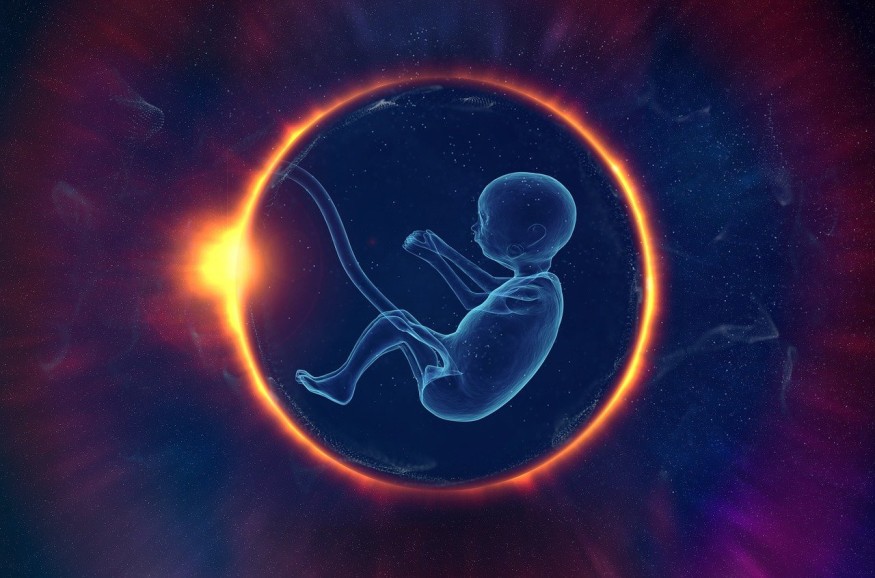
Lab-Grown Human Embryos
The embryo models hold vital features that have not been observed in earlier models. These include three different lineages that facilitate embryonic support structures as well as the placenta. It also includes cell layers that form into an embryo prior to folding and that grow into various organs and tissues.
Earlier research revealed that stem cells taken from embryos of mice could still be guided artificially for them to grow into tissues that make up and support the embryos themselves. In such a case, they assemble on their own to form a SEM at the stage of post-gastrulation, wherein cells of the embryo turn into three primary body tissue types.
Hanna and the team explain in their study that they extend such findings to a human context with the help of genetically unmodified human embryonic stem cells.
The researchers pinpointed ideal conditions, such as cell mixture ratios and cell numbers, starting from implantation that takes place roughly seven to eight days post-fertilization. As such, they explain that the full SEMs exhibited dynamics in developmental growth that mirror the hallmarks of embryogenesis' post-implantation phase around 13 to 14 days after fertilization.
The embryonic models show the mix of all lineages that are known and all components of human embryos in the early stage. These include the hypoblast, epiblast, trophoblast, yolk sac, and extraembryonic mesoderm.
The SEM dataset's cell profiles were also seen to mirror cell type composition and genetic expression patterns among human embryos shortly post-implantation, when compared to a benchmarking dataset.
While the SEMs are not strictly identical with human embryos, they serve as a model that leads to various possibilities in research.
Hanna explains that their models can be utilized to shed light on the mechanical and biochemical signals that make sure that proper development takes place during the early stage of pregnancy and on areas where development could end up wrong.
Check out more news and information on Medicine and Health in Science Times.
© 2026 ScienceTimes.com All rights reserved. Do not reproduce without permission. The window to the world of Science Times.


