Swiss Federal Institute of Technology Lausanne EPFL experts developed a new imaging approach through the combination of two distinct microscopy techniques. The assembly of the device is part of a study to observe the various processes of a cell on its membrane. The authors believe that through the examination of cells, we would be able to identify the simultaneous cellular progression in many human body conditions. In addition, the study could give insights as to what happens to the cells during an infection.
Importance of Studying Cells
Cells are among the biological materials that contain the essential roles of any living species. There are many types of cells, with each taking a complex process to achieve their specified functions. Studying their structures, how they move, and what impacts they have are essential for experts to identify detailed information during disorders and diseases and improve the treatments available today. However, the potential of the findings that could be gathered from the studies is challenging to achieve due to the difficulty of observations from the micro and nano scales of the cells.
To solve the problem of getting images from cells, researchers from the EPFL and experts from two separate laboratories conducted a collaborative effort to develop a new kind of imaging that can observe the tiny cells from living organisms.
EPFL Laboratory for Bio- and Nano-Instrumentation, Institute of Bioengineering expert and author of the study Georg Fantner said in a PhysOrg report that the granular properties make it harder for many studies to observe the cells, and even the modern-day technologies are still unable to procure a stabilized imaging results.
Combining Electron and Fluorescence Microscopy for a Better Cell Observation
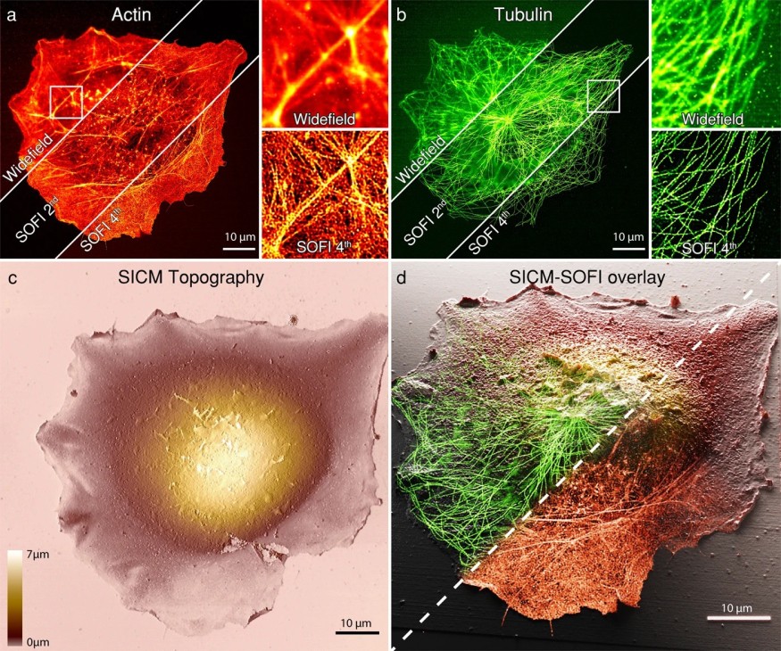
According to the expert, electron microscopy is among the most advanced and best solutions to observe the cell surface and nanoscale structure. However, electron microscopy requires a complex process in which the specimens are placed in a vacuum while being targeted with plenty of electrons. This phase would not allow any living specimens, or in this case, the cells.
Alongside the electron-bombarding image technique, fluorescence microscopy holds a promising effectivity to view the cellular bodies. Unlike electron microscopy, this approach does not have any capacity to destroy the subject but to perform the full resolution of the imaging; fluorescence microscopy requires a complex and seemingly impossible configuration. By notching up the image's resolution from the subject, photons are also heightened, which could exceed its limits and eventually inflict damage to the specimen.
The capacity of both electron and fluorescence microscopy is unparalleled but is also hindered by the limitations above the potential it offers. With that said, the experts concluded to combine the beneficial factors of both the technologies in a single device to materialize a novel approach to cell imaging. With the development, numerous studies will go through convenient cell examination without any evasive methods toward the subjects.
The studies were published in the journal ACS Nano, titled "Time-Resolved Scanning Ion Conductance Microscopy for Three-Dimensional Tracking of Nanoscale Cell Surface Dynamics," and in Nature Communications, titled "Correlative 3D microscopy of single cells using super-resolution and scanning ion-conductance microscopy."
RELATED ARTICLE : Microscopic Robot Inspired From Starfish Larva Moves Around Using Tiny Hairs to Deliver Medicine to Cells
Check out more news and information on Nanotechnology in Science Times.
© 2026 ScienceTimes.com All rights reserved. Do not reproduce without permission. The window to the world of Science Times.


